
Frequent Power Change
Spectacle power may change repeatedly as corneal shape becomes more irregular.
See corneal mapping →Keratoconus can cause frequent power changes, irregular astigmatism, ghost images, glare and progressive blur. SGVEH supports keratoconus evaluation with corneal imaging, specialist assessment and treatment planning including C3R/CXL, visual rehabilitation and advanced corneal care when needed.
Keratoconus affects the shape and strength of the cornea. Symptoms and treatment needs vary widely, so a cornea-focused evaluation is important before deciding what to do next.
Spectacle power may change repeatedly as corneal shape becomes more irregular.
See corneal mapping →Irregular corneal shape can create multiple outlines, halos or shadowed images.
Ask for cornea evaluation →Vision may remain unclear even with glasses when astigmatism becomes irregular.
Read common questions →Young patients may need monitoring to see whether the cornea is becoming steeper or thinner.
See care pathway →Cross-linking may be considered when suitable keratoconus is documented to be progressive.
Treatment options →RGP, hybrid or scleral lens strategies may help selected patients achieve better functional vision.
Visual rehabilitation →Advanced keratoconus may produce scarring and substantial reduction in corrected vision.
Need advanced cornea care? →Corneal transplant is generally reserved for selected advanced cases when other options are insufficient.
Know when transplant is discussed →For a keratoconus-focused page, the cornea specialist comes first. Supporting comprehensive and refractive-care doctors follow, with the wider SGVEH multispecialty team available when broader ophthalmic care is needed.

MBBS, DOMS; advanced cornea training
Cornea • Keratoconus • Ocular Surface • Corneal Transplant

MBBS, MS (AIIMS New Delhi)
Cataract • Refractive Care • Glaucoma • Neuro-Ophthalmology

Ophthalmologist
Strategic leadership, hospital systems and comprehensive eye care.

Ophthalmologist
Comprehensive ophthalmology and patient care with coordinated follow-up support.

Ophthalmologist
Comprehensive ophthalmology and patient care support.

Ophthalmologist
Comprehensive ophthalmology and clinical care within the SGVEH network.

Ophthalmologist
Holistic eye care, community ophthalmology and institutional direction.

Ophthalmologist • Retina Specialist
Medical and surgical retina support within the wider multispecialty eye-care network.

Ophthalmologist • Retina Specialist
Retina, uvea and vitreoretinal care within the SGVEH specialist network.

Ophthalmologist
Children’s eye care, amblyopia and squint care for pediatric ophthalmic needs.

MBBS, MS, FACS; fellowship training
Oculoplasty, ptosis, DCR, orbit and eyelid reconstruction support.
Treatment suitability depends on corneal shape, thickness, progression, scarring, age and visual needs. Keratoconus care should be planned after an in-person corneal examination and appropriate imaging.
Because keratoconus changes the shape and thickness of the cornea, imaging is important for diagnosis, staging and monitoring progression.
Maps corneal curvature and helps identify irregular steepening patterns.
Provides detailed anterior and posterior corneal shape and thickness information when clinically indicated.
Pachymetry helps assess how thin the cornea is and whether treatment options are suitable.
Measures how much vision can be improved with glasses or lenses and how irregular the optics have become.
Looks for corneal signs, scarring and surface-related issues that may affect treatment planning.
Serial scans help determine whether the cornea is changing over time and whether stabilization treatment should be discussed.
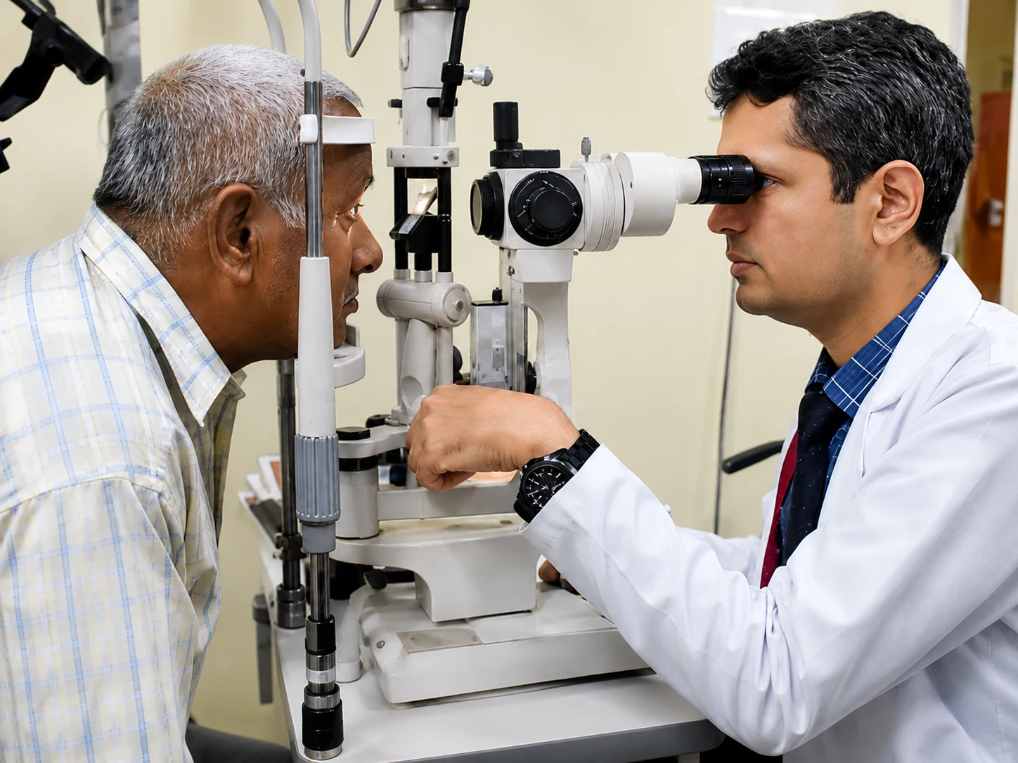
The goal is to understand both visual quality and whether the cornea is progressing before choosing treatment.
Understand symptoms, age, spectacle changes, rubbing history and previous corneal scans if available.
Map the cornea and assess thickness, irregularity, scarring and evidence of progression.
Discuss visual correction and stabilization options such as specialty lenses or C3R/CXL when clinically suitable.
Continue follow-up with repeat imaging to monitor stability and guide advanced care if needed.
Some patients need better visual correction, some need corneal stabilization, and advanced cases may need surgical corneal options. The treatment plan should match disease stage, progression and functional vision.
Simple answers to common questions patients ask before consulting a cornea specialist.
Need personalised guidance? →Keratoconus is a condition in which the cornea becomes thin and develops an irregular cone-like shape. This can cause blurred vision, astigmatism, glare and visual distortion.
C3R/CXL is a corneal strengthening treatment used in selected progressive keratoconus. Its main purpose is generally to reduce further progression rather than to guarantee spectacle-free vision.
Yes, many patients do not need transplant. Depending on the stage, management can include glasses, specialty contact lenses, cross-linking and other cornea procedures.
Standard LASIK is generally not suitable in established keratoconus because the cornea is structurally weak. A cornea specialist should assess refractive options individually.
Pentacam/tomography can help map corneal shape and thickness, detect subtle changes and compare progression over time.
Transplant may be discussed in selected advanced cases with significant scarring, very poor corrected vision or when other measures are insufficient. Suitability requires specialist examination.
This page is educational. Keratoconus treatment depends on corneal imaging, progression, thickness, visual function and clinician assessment. An in-person cornea examination is required before deciding whether C3R/CXL, specialty lenses or surgery is appropriate.
Our team can help guide you toward an appropriate SGVEH keratoconus and cornea evaluation pathway in Raipur.